DEFINITION
An aneurysm is defined as a permanent abnormal dilatation of a blood vessel occurring due to congenital or acquired weakening or destruction of the vessel wall. Most commonly, aneurysms involve large elastic arteries, especially the aorta and its major branches. Aneurysms can cause various ill effects such as thrombosis and thromboembolism, alteration in the flow of blood, rupture of the vessel and compression
of neighbouring structures.
CLASSIFICATION
Aneurysms can be classified on the basis of various features:
A. Depending upon the composition of the wall:-
1 True aneurysm composed of all the layers of a normal vessel wall.
2. False aneurysm having fibrous wall and occurring often from trauma to the vessel.
B. Depending upon the shape:-
1. Saccular having large spherical outpouching.
2. Fusiform having slow spindle-shaped dilatation.
3. Cylindrical with a continuous parallel dilatation.
4. Serpentine or varicose which has tortuous dilatation of the5. Racemose or circoid having mass of intercommunicating
small arteries and veins.
C. Based on pathogenetic mechanisms:-
1. Atherosclerotic (arteriosclerotic) aneurysms are the most common type.
2. Syphilitic (luetic) aneurysms found in the tertiary stage of the syphilis.
3. Dissecting aneurysms (Dissecting haematoma) in which the blood enters the separated or dissected wall of the vessel.
4. Mycotic aneurysms which result from weakening of the arterial wall by microbial infection.
5. Berry aneurysms which are small dilatations especially affecting the circle of Willis in the base of the brain
Three common type of aortic aneurysm.
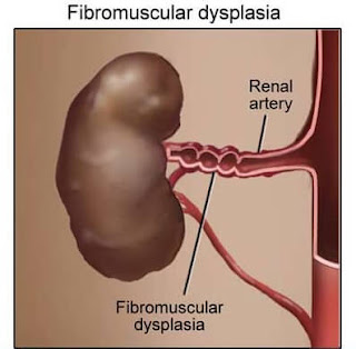
FIBROMUSCULAR DYSPLASIA
Fibromuscular dysplasia first described in 1976, is a non atherosclerotic and non-inflammatory disease affecting arterial wall, most often renal artery. Though the process may involve intima, media or adventitia, medial fibroplasia is the most common.
MORPHOLOGIC FEATURES.
Grossly, the involvement is characteristically segmental—affecting vessel in a bead like pattern with intervening uninvolved areas.
Microscopically, the beaded areas show collections of smooth muscle cells and connective tissue. There is oftenrupture and retraction of internal elastic lamina.
The main effects of renal fibromuscular dysplasia,depending upon the region of involvement, are renovascular hypertension and changes of renal atrophy.


ConversionConversion EmoticonEmoticon